פרופ’ דניאל קורניצר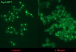
המחלקה למיקרוביולוגיה מולקולרית
הבסיס המולקולרי לאלימות של פטריות פתוגניות
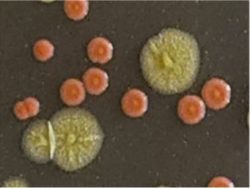
זיהומים הנגרמים מפטריות פתוגניות נהיים שכיחים יותר, בד בבד עם גידול האוכלוסיות הרגישות. Candida albicans הוא הפתוגן הפטרייתי הסיסטמי הנפוץ ביותר. Candida auris הוא פתוגן מתעורר, אשר שמתפשט בהדרגה בכל העולם, והוא עמיד למרבית הטיפולים. המעבדה חוקרת מנגנונים המאפשרים לפטריות אלה לשגשג במאכסן האנושי, כגון מערכות לקליטת ברזל מהמוגלובין (ראו תמונה).
פרויקטים מוצעים מתבססים על מערכת שפותחה לאחרונה לאנליזה גנטית של פטריות אלה, כדי לנתח את המסלולים התאיים שמעורבים באלימותן.
כתובת מייל: danielk@technion.ac.il
פרופ’ עמי אהרונהיים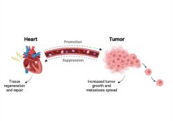
מחלקה: ביולוגיה של התא ומדעי הסרטן
אי ספיקת לב וסרטן הם בין גורמי התמותה המובילים בעולם המערבי. הלב מגיב ללחצים חריפים שונים על ידי תהליכי בנייה אדפטיביים; עם זאת, כאשר הלחץ הופך לכרוני, השינויים הללו הופכים לבלתי מסתגלים, מה שגורם לגדילה לא מבוקרת של הלב, התפתחות תהליכי צלקת המובילים לאי ספיקת לב. מחלות לב וסרטן חולקים גורמי סיכון דומים, כולל סכנות סביבתיות, נטייה גנטית, עישון, השמנת יתר, אורח חיים בטלני, סוכרת והזדקנות. גדילה לא מבוקרת של שריר הלב נחשבת זה מכבר לתהליך הדומה לגידול סרטני. במעבדתי שילבנו את שתי המחלות במספר מודלים בעכברים. מצאנו שהתקדמות תהליך סרטני משפרת את תפקוד התכווצות הלב, מפחיתה גדילה לבבית לא מבוקרת, ותהליכי בניית צלקת. האם ייתכן שסרטן יכול לשמש דרך טיפולית מבטיחה לאי ספיקת לב? טיפול באי ספיקת לב באמצעות גידול סרטני אינו אופציה לטיפול. עם זאת, האם ניתן לרתום את התהליכים סרטן לטיפול באי ספיקת לב? במעבדתנו פיתחנו פלטפורמה לחקור את המנגנונים המולקולריים המעורבים בתהליך זה תהליכים שעשויים להוביל לאפיקים טיפוליים חדשים לשיפור מצבו של לב כושל ומחלות המערבות צלקת ואף מחלות אחרות.
כתובת מייל:aronheim@technion.ac.il
 פרופ’ משה גביש
מחלקה: נוירוביולוגיה
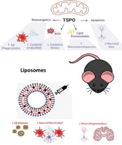
תפקיד החלבון TSPO במצבי בריאות וחולי.
החלבון TSPO ( Translocator protein) ממוקם בממברנה החיצונית של המיטוכונדריה ויש לו תפקיד חשוב בקליטה של כולסטרול למיטוכונדריה, יצירת סטרואידים, פרוליפרציה של תאים, נשימה תאית, תהליכי מוות תאי מתוכנת ופעילות של תאי מערכת החיסון. בשנים האחרונות המעבדה מתמקדת בתפקיד של TSPO בתהליכי דלקת ואפשרות להשתמש בליגנדים לחלבון זה כטיפול במחלות דלקתיות. נמצא כי לחלבון יש תפקיד מרכזי בוויסות של פעילות פגוציטים ותאי מיקרוגלייה שהם התאים המרכזיים בוויסות תגובה דלקתית היקפית ומוחית. המעבדה פיתחה ליגנדים חדשים לחלבון זה ומצאה שלליגנדים אלו יש השפעה אנטי- דלקתית במוח ובאיברים היקפיים. נמצא כי אחד הליגנדים מעכב את פעילות החלבון NFK-B שהוא חלבון המפתח ביצירת ציטוקינים פרו-דלקתיים. השפעה אנטי-דלקתית זו נצפתה גם במחקרים ברמה התאית-מולקולרית וגם במודלים של דלקת במכרסמים. הליגנד הדגים השפעה תראפויטית במודלים למחלת פרקינסון, דלקת מוחית, דלקת מעי, טראומה מוחית ודלקת מוחית אוטואימונית. המעבדה פיתחה מערכת שחרור מושהה המבוססת על ליפוזומים והדגמנו יכולת להיטיב מחלת פרקינסון שמושרית במכרסמים וביונקים. הליפוזומים שפותחו לצורך מעבר דרך מחסום דם-מוח כללו פפטיד ייחודי שמכוון את הליפוזומים לטרנספורטר במוח. מערכת זו שימשה גם לטיפול במודל גליובלסטומה ודלקת מוחית.
כתובת מייל: mgavish@technion.ac.il
 פרופ”ח רובי שלום-פוירשטיין
מחלקה: גנטיקה וביולוגיה התפתחותית
המעבדה לחקר תאי גזע:
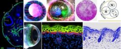
תאי גזע מחדשים את הרקמות שלנו בשגרה או לאחר פציעה. למרות שנים של מחקר, תכונותיהם הבסיסיות של תאי הגזע עדיין אינן מובנות היטב. לדוגמה, לא ברור כמה תאי גזע קיימים, מה הגנים המעורבים בבקרת הפעולה של תאי הגזע, וכיצד הם מווסתים על ידי הסביבה שלהם בבריאות ובחולי.
במעבדה ביססנו מערכת המאפשרת מעקב חי אחר תאי הגזע והצאצאים שלהם בקרנית ובעור של עכברים טרנסגניים. מערכת ייחודית זו חושפת את הדינמיקה של התחדשות הרקמה על ידי תאי הגזע, האינטראקציות שלהם עם הסביבה המקומית שלהם, וכשל בפעולת תאי גזע במחלות. בפרויקט המוצע, נחקור את המנגנונים המולקולריים שמאחורי תפקוד תקין או פגום של תאי גזע ונבחן את מנגנון התחדשות של אוכלוסיות תאי הגזע לאחר אובדן מוחלט שלהם.
כתובת מייל: SHALOMFE@TECHNION.AC.IL
מנגנוני פירוק של רנ״א וחלבונים בעקה
מעבדה של ד״ר בוריס סלובודין, המחלקה לביוכימיה
אז מה אנחנו חוקרים? אנחנו רוצים לגלות איך תאים אנושיים שורדים מצבים של סטרס. כדי להבין זאת, אנחנו חוקרים את מנגנוני הפירוק של מולקולות רנ״א וחלבונים במצבים של נזק ל-DNA התאי. למה זה חשוב? מצבי עקה נפוצים מאוד בחיי התא האנושי וחשוב מאוד להבין את דרכי ההתמודדות איתם שהתפתחו במהלך האבולוציה. האם זה קשור למחלות? בהחלט, מצבי עקה קשורים למגוון מחלות והרבה מהן פורצות עקב יכולת מוגבלת להתמודד עם מצבם אלו. למשל, מחלת הסרטן הרבה פעמים נובעת מיכולת מוגבלת לתקן נזקים ב-DNA התאי. באילו שיטות אנחנו משתמשים? אנחנו משתמשים במגוון שיטות, כולל ביוכימיות קלסיות, גנומיות, גנטיות, רובוטיות, ומבוססות בינה מלאכותית. אנחנו גם מרצפים את כלל הגנים בשיטות של ״ריצוף עמוק״ ובמקביל משתמשים במיקרוסקופים אוטומטיים כדי לאסוף נתונים באופן רציף. באיזה מודלים מתבצע המחקר? רוב המחקר מתבצע בתאי אדם בתרבית ולאחרונה אנחנו גם משתמשים בספרואידים. מה זה ספרואידים? אלו תאים שגודלו בתנאים מיוחדים שגורמים לתאים ליצור מבנה כדורי. האם תעבוד/י לבדך? לא, העבודה מתבצעת בצוות, בהדרכה של סטודנט/ית מנוסה יותר, בד״כ דוקטורנט/ית. האם תצטרך/י לעבוד כל הזמן תחת השגחה? בהחלט לא, לאחר תקופת הכשרה, ניתן יהיה לעבוד באופן עצמאי יותר. האם תוכל/י להשתתף בתכנון הניסויים? בהחלט, עם הזמן תוכל/י לתכנן ניסויים בעצמך. מה תשיג/י בתקופת הפרוייקט? במהלך הפרוייקט תלמד/י לעבוד במעבדה, לתכנן ניסויים באופן נכון, לבצע אותם באופן מדויק ולהסיק מסקנות. חשוב מאוד, תוכל/י להבין האם המחקר המדעי ״מדבר אליך״ והאם תרצה/י לעסוק בו בעתיד. האם העבודה היא בשעות גמישות? בהחלט, הסטודנטים יכולים לבחור את שעות עבודתם בתיאום עם המדריכ/ה. בתקופות לחוצות יותר, כגון תקופת בחינות, אפשר לעבוד פחות. ואם תגלה/י משהו חשוב, האם שמך ייכלל ברשימת כותבי המאמר? בהחלט, מדיניות המעבדה היא שכל מי שתורם תרומה משמעותית למחקר כלשהו, נכלל ברשימת כותבי המאמר הרלוונטי. איך תוכל/י לבדוק פרטים נוספים וליצור קשר? אפשר ללחוץ על הלינק שמוביל לאתר המעבדה. שם ניתן לקרוא עוד פרטים על המחקר המתבצע, להכיר את צוות המעבדה, להציץ לחיי החברה וגם להגיש מועמדות לפרוייקט. בהצלחה בהמשך!
https://slobodin.net.technion.ac.il
 ד”ר אורן כספי
מחלקה: פיזיולוגיה
מנהלת המעבדה: ד”ר חווה ליכטיג-קינסבורנר
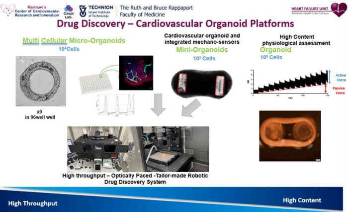
תחומי העניין של המעבדה: המעבדה עוסקת באורגנואידים (אברונים) לבבים רב-תאיים המכילים את כל מגוון התאים בלב האנושי (קרדיומיוציטים, אנדותל, פיברובלסטים לבביים, ותאי שריר חלק ומערכת החיסון). המעבדה מפתחת מודלים מעבדתיים למחלות לב גנטיות ונרכשות. לאחרונה המעבדה פיתחה מודל פורץ דרך, ראשון מסוגו בעולם, לאי ספיקת לב עם מקטע פליטה שמור HFpEF. המעבדה עוסקת הן בטכנולוגיות לפיתוח מודלים משופרים לגילוי תרופות וסקירת אפקטים טוקסיים והן בבדיקת טיפולים ומנגנוני טיפול אפשריים על בסיס המודלים שפותחו.
הפרויקטים המוצעים:
פרויקט חישובי – שימוש בשיטות בינה מלאכותית לשיפור ההדירות (reproducibility) ואיכות המדדים הפיזיולוגים הנמדדים באורגנואידים רב-תאיים.
פרויקט מולקולרי – שימוש בכלים מולקולריים מבוססי CRISPR/Cas9 להשפעה על פיברוזיס במהלך ההתפתחות של אי ספיקת הלב.
כתובת מייל: caspio@technion.ac.il
כתובת אתר המעבדה: www.caspilab.com
הבסיס העצבי של למידה והתנהגות חברתית
מחלקה: נוירוביולוגיה
ד”ר בן אנגלהרד
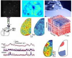
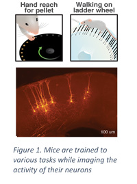
במעבדה אנחנו מנסים להבין איך פעילות של תאי עצב במוח מאפשרת התנהגויות מורכבות כגון למידה והתנהגות חברתית. אנחנו משתמשים בכלים מתקדמים כגון דימות פונקציונלי ברמה של תא בודד בחיות מתנהגות, מיקרוסקופיית מולטיפוטון, כלים חישוביים ואלגוריתמי למידה עמוקה, מערכות מציאות מדומה לעכברים, מודלים של עכברים עם הפרעות כגון אוטיזם, ועוד. סטודנטים במעבדה נחשפים למגוון כלים בחזית של חקר המוח בסביבה קולגיאלית ונעימה.
 הפרויקט המוצע: במעבדה יש מגוון פרויקטים שניתן לבחור:
הקמת מערכת לבדיקת הבסיס העצבי של קבלת החלטות חברתיות. בפרויקט הסטודנטית תקים ותבדוק מערכת חדשה המאפשרת לעכבר להחליט בן קבלת תגמול לא חברתי (למשל אוכל) לבין קבלת אינטרקציה חברתית, ובין קיום אינטראקציות חברתיות שונות (למשל בין עכבר מוכר לעכבר חדש). המערכת תתוכנן בתוכנה של תלת מימד ותורכב ע”י הסטודנט. לאחר הקמת המערכת היא תבחן ע”י שימוש בעכברים. אם יתאפשר הזמן נבדוק גם עכברי מודל של הפרעות שונות.
הקמת מערכת לבדיקת הבסיס העצבי של אינדיבידואליות. בפרויקט הסטודנטית תקים ותבדוק מערכת חדשה המאפשרת לעכבר לבנות את אזור המחסה שלו בדרכים שונות. מה שונה בפעילות העצבית של עכברים העושים פעולה זו בצורה שונה? המערכת תתוכנן בתוכנה של תלת מימד ותורכב ע”י הסטודנט. לאחר הקמת המערכת היא תבחן ע”י שימוש בעכברים. אם יתאפשר הזמן נבדוק גם עכברי מודל של הפרעות שונות.
עיבוד תמונה של פעילות מוחית. זהו פרויקט חישובי בו הסטודנטית תיישם אלגוריתם של עיבוד תמונה בסרטים אשר נלקחו ממיקרוסקופ הבוחן פעילות מוחית. האלגוריתם מאפשר תיקון של תנועות לא אחידות שקורות במהלך הרישום. הדגש הוא על יישום יעיל של האלגוריתם. ידע קודם בתכנות עם GPU – יתרון.
נא לשלוח למייל לבן אנגלהרד: benengelhard@gmail.com
אתר: Engelhard Lab – Neural circuits for Complex Behavior (technion.ac.il)
 ביולוגיה של תנועות הפנים ביונקים
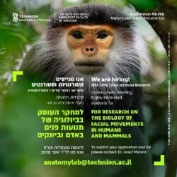
ד״ר אסף מרום
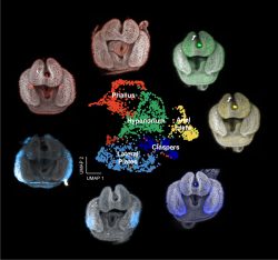
תנועות הפנים של יונקים מאפשרות תפקודים חיוניים כמו הגנה על העין, שמיעה והזנה. חלק מתנועות אלה משמשות גם להבעות – קומפלכס של תנועות אשר יש להן מִתאם עם מצב רגשי מסויים, למשל: עצב, או שמחה. באדם, הבעות הפנים מגוייסות גם לצורך תפקוד מיוחד – דיבור. דרווין היה זה שטען שהבעות הפנים של האדם הן אוניברסליות, ושהן התפתחו מתנועות פנים דומות אצל בעלי חיים אחרים.
מטרת המעבדה להשיג הבנה מנגנונית של תנועות הפנים. לפיכך, הפרוייקטים מרוכזים סביב שלוש שאלות מרכזיות: מהי הארכיטקטורה של שרירי הפנים ביונקים אשר מאפשרת את התנועות? כיצד שרירים אלה נשלטים על ידי מערכת העצבים המרכזית? וכיצד מתאפשרת הקליטה והפרשנות של הבעות הפנים על ידי הזולת? בנוסף לניסיון ללמוד את תנועות הפנים הנורמליות, נבחן תנועות אלה גם במצבי חולי.
סטודנטים/ות מוזמנים/ות לפנות בדוא״ל: assafma@technion.ac.il או anatomylab@technion.ac.il
 התחזוקה וגדילה של סרקומרים בלב הבריא והחולה
פרופ’ יצחק קהת
מחלקה: פיזיולוגיה
תחומי העניין של המעבדה: בכל תא לב יש כ 1000 מנועים הקרויים סרקומרים, ובלב כולו 2-3 טריליון סרקומרים. תאי הלב מחליפים את כל חלקי המנועים (החלבונים) כל מספר ימים, בזמן שהמנועים עובדים ברציפות. אנחנו חוקרים איך הלב יכול להחליף את כל החלקים של 2-3 טריליון מנועים תוך כדי שהם עובדים, ואיך כשלים במערכת תורמים להתפתחות אי ספיקת לב.
פרויקט המוצע: פיתוח שיטות חדשניות על מנת לזהות את המרכיבים של מערכת החלפת החלבונים בסרקומר כדי להבין איך המערכת עובדת , מתי ולמה היא כושלת, ואיך ניתן למנוע זאת.
כתובת מייל: ikehat@technion.ac.il
מערכת האוביקיטין לפרוק חלבונים ומעורבותה בממאירויות ובמחלות ניווניות של מע’ העצבים המרכזית
אהרן צ’חנובר, MD, DSc – פרס נובל לכימיה לשנת 2004
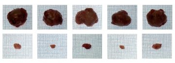
הסבר לציור: כשנועלים את מערכת האוביקיטין בגרעין באמצעות תרמית מטבולית – ניתן להרעיב גידולים סרטניים למוות. האפקט הוא סלקטיבי לתאים ממאירים, כי דרישתם המטבולית לחומצות אמיניות המסופקות על ידי מערכת האוביקיטין גדולה משל תאים בריאים, ולכן נעילה חלקית של המערכת בגרעין תפגע בהם באופן סלקטיבי. בשורה העליונה – גידולים בעכברים שלא טופלו, ובשורה התחתונה – גידולים בעכברים שטופלו.
המרכז הבינ-תחומי לחקר הסרטן על שם ע”ש רפפורט
המעבדה עוסקת בעיקרה במנגנונים בסיסיים המקשרים בין מערכת האוביקיטין לפרוק חלבונים (פרס נובל לכימיה לשנת 2004) שהתגלתה על ידי הפרופסורים הרשקו, צ’חנובר ורוז, לפתוגנזה של מחלות – ביניהן ממאירויות ומחלות ניווניות של מע’ העצבים המרכזית. פענוח מעט מהמנגנונים מאפר לנו המעבדה כבר עתה, לנסות ולפתח תרופות, אחת מהן נמצאת כבר בשלב מסחרי מתקדם.
הבנת המנגנון מדכא ההתמרה של עודף במרכיב p50 של פקטור השעתוק NF-kappaB או מנגנוני הפרוק של חלבונים המעורבים בפתולוגיה של Amyotrophic Lateral sclerosis – ALS.
כתובת מייל: aaroncie@technion.ac.il; meiravf@technion.ac.il
פענוח מנגנון פעולתן של מולקולת גזיות ביולוגיות בתאים סרטניים
מעבדה של פרופ’ מורן בנהר
ביוכימיה
תחומי העניין של המעבדה
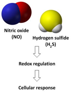
פענוח מנגנון פעולתן של מולקולת גזיות ביולוגיות בתאים סרטניים .
תהליכי חמצון-חיזור ביולוגיים, בפרט השפעת ניטריק אוקסיד
ומימן גופריתי על מסלולי דלקת ומוות תאי- בתאים
מקרופאגים ותאים סרטניים. הפרויקט המוצע: מציאת חלבוני מטרה
של ניטריק אוקסיד ומימן גופריתי בתאי סרטן בתגובה לטיפולים אנטי-סרטניים
benhar@technion.ac.il
 המעבדה לחקר העין
פרופ ניצה גולדנברג כהן , מנהלת מחלקת עינים, ביח בני ציון.
מחלקה: עיניים, הפקולטה לרפואה , קומה 13
תחומי העניין של המעבדה:
המעבדה עוסקת במחקר תרגומי מהקליניקה למעבדה
ובחזרה. מודלים של מחלות עינים, בעיקר של עצב
הראיה, הכולל גלאוקומה, נזק אסכמי של עצב הראיה, נזק
דלקתי של עצב הראיה, מודל סכרת, קטרקט , פרוטזות ועוד. כמו כן שמוש בטכנולוגית נאנו, בדיקת טוקסיות
של מתכות בדמעות וברקמות .
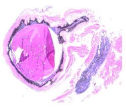
הפרויקט המוצע: מוצעים מגוון פרויקטים , הכוללים עבודה עם מודל עכבר בעיקר בשלב חיתוך הסטולוגי ובצוע
צביעות אימונו- הסטוכימיות של עינים של עכבר אחרי השריית נזק, צלום וחישוב סטטיסטי של התוצאות,
בדיקות מולקולריות לבטוי גנטים, דגימות מבני אדם וממודל עכבר.
כתובת מייל :
Ncohen1@gmail.com
Nitza.Cohen@b-zion.org.il
 שחזור העולם החיצוני מתוך פעילות המוח
פרופ’ דורי דרדיקמן
מחלקה: חקר המוח
תחומי העניין של המעבדה: איך עובד הזכרון במוח? אחד מסוגי הזיכרון החשובים ביותר הוא הזכרון האסוציאטיבי. דוגמא אחת לזכרון אסוציאטיבי היא זכרון של מקומות – כאשר אנחנו נמצאים במקום מסויים, עולים בנו זכרונות הקשורים למקום שאנו נמצאים בו. אנו חוקרים את הזכרונות הללו, תלויי המקום. אנו משתמשים בטכנולוגיות של רישום וגירוי של תאי מוח רבים בו-זמנית, מתוך מטרה לקשר בין הפעילות המוחית הזאת לבין השאלה: איך אפשר לזכור איפה אנחנו נמצאים? מה ההשלכות של זכרון זה?
הפרויקט המוצע: תאי מקום בהיפוקמפוס הם חלק מהייצוג הפנימי במוח של הסביבה בה אנו נמצאים. יש ברובוטיקה בעייה ידועה שקוראים לה בעיית ה-SLAM, או Simultaneous Localization and Mapping. השאלה הזאת רלוונטית גם למוח ולא רק לרובוטיקה – איך אנחנו יכולים לייצר ייצוג של מפת הסביבה ובמקביל לייצר ייצוג של המקום שלנו בתוכו. ידוע כבר שנים רבות שניתן לשחזר את המקום של העכבר מתוך פעילות תאי המקום שלו. מאידך, הבעייה ההפוכה לא פתורה – האם ניתן לשחזר את מפת הסביבה מתוך הפעילות המוחית? זו בדיוק מטרת הפרוייקט הזה – אנו נרשום פעילות של מאות תאי מוח במקביל בזמן שעכברים מנווטים בסביבות מורכבות, וננסה לשחזר את מפת הסביבה מתוך הפעילות המוחית. זה יאפשר לנו להבין נדבך חשוב נוסף בקשר בין המוח לעולם שסביבו.
כתובת מייל: פרופ’ דורי דרדיקמן derdik@technion.ac.il
מחקר בנושא הבסיס של הגנטי של מחלות נדירות
ד”ר קארין ויס
מחלקה: מכון גנטי רמב”ם
תחומי העניין של המעבדה: איתור הגורם הגנטי למחלות נדירות נוירו-התפתחותיות לא מאובחנת ושיפור המעקב והטיפול בחולים.
הפרויקט המוצע: השתלבות בפרויקטים העוסקים בהבנת הגורם הגנטי בחולים עם מחלות נדירות כולל בדיקות גנטיות חדשניות, ביואינפורמטיקה, אנליזה של רנא וחלבון בתאי החולה, יצירת מודל חיה ועוד.
כתובת מייל:k_weiss@rmc.gov.il
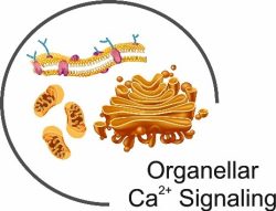 תפקידם של תעלות סידן בבקרת מערכת החיסון
ד”ר רז פלטי
מחלקה: ביוכימיה
מעבדת המחקר מתמחה במנגנוני העברת סיגנל תאיים המעורבים בהתפתחות סרטנית ובפעילות מערכת החיסון. אנו חוקרים כיצד אורגנלות תוך תאיות מתקשרות על מנת לווסת את עוצמת, משך ודפוס סיגנל הסידן התוך תאי וכיצד פעילות זו משתתפת בתהליכים סרטניים ובמחלות של חסר-חיסוני. בנוסף, המעבדה עוסקת בפיתוח שיטות חדשניות המבוססות על פוטו-פרמקולוגיה ופוטו-גנטיקה לשליטה על פעילות אנטי סרטנית של מערכת החיסון.
הפרויקט המוצע: פעילותם של חלבוניSTIM1 ו- ORAI1 נחוצה לשפעול של לימפוציטים באדם ומוטציות שונות בגנים המקודדים לחלבונים אלו עומדות בבסיס סינדרום נדיר של חסר-חיסוני קשה. הפרויקט יעסוק בהבנת המנגנון שדרכו שני החלבונים מתקשרים זה עם זה על מנת ליצור אותות סידן בלימפוציטים.
כתובת מייל:razpalty@technion.ac.il
מנגנוני התחמקות ממוות ע”י תאי סרטן
פרופ”ח חגי וולפזון
מחלקה: גנטיקה וביולוגיה התפתחותית
מעבדת וולפנזון חוקרת את האינטראקציות של תאים עם המטריקס החוץ-תאי, את ההחלטות שמתקבלות ע”י תאים כתגובה לסיגנלים שמתקבלים מהמטריקס, ואת המנגנונים שגורמים לשיבוש של החלטות אלו. אחת הדוגמאות המרכזיות ביותר נוגעת ליכולת של תאי סרטן לשרוד ולגדול בסביבות מגוונות מאוד ללא קשר לסיגנלים מהמטריקס, בעוד שתאים לא סרטניים עוברים מוות תאי (אפופטוזיס) כאשר הם נמצאים בסביבה שלא מאפשרת היקשרות למטריקס. כחלק מהפרויקט, הסטודנטית/סטודנט תיקח/יקח חלק בחקר המנגנון שדרכו תאים סרטניים מתחמקים ממוות תאי ובמציאת דרכים לשינוי ההחלטה התאית הזו. הפרויקט יכלול עבודה עם תאים, מיקרוסקופיה מתקדמת, אנליזות ממוחשבות ועבודה מולקולרית.
למתעניינים, אנא לפנות לפרופ”ח חגי וולפנזון: haguyw@technion.ac.il
רשתות בקרה של הומאוסטזיס חלבונים בסטרס ובמחלות נוירודגנרטיביות
פרופ״ח רעות שלגי
מחלקה: ביוכימיה
מעבדת שלגי חוקרת בקרות של הומאוסטזיס חלבונים בתאי יונקים, בגישה של ביולוגיה מערכתית (systems biology), המשלבת כלים מולקולארים, חישוביים, סריקות וטכנולוגיות כלל גנומיות חדישות, כדי להבין לעומק
רשתות בקרה ברמת השיעתוק, splicing, תרגום, וקיפול חלבונים. אנו שואלים/ות כיצד רשתות אלו מותאמות לאפשר אדפטציה תאית לתנאים של סטרס, ומה משתבש במחלות נוירודגנרטיביות.
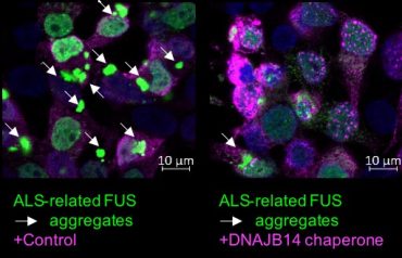
לאחרונה זיהינו במעבדה מנגנונים ייחודיים לסוג של ALS הנגרם על ידי אגרגציה של החלבון FUS, והצלחנו לאפיין חלבונים המסוגלים למנוע כמעט לחלוטין את האגרגציה. זהו צעד ראשון בדרך לזיהוי הכלים הספציפיים המאפשרים לנו להילחם במחלה זו.
לאתר המעבדה: https://reuts4.wixsite.com/reutshalgi
הפרויקט המוצע:
מעבדתנו מציעה שני סוגים של פרויקטים:
בקרת ביטוי גנים במחלת ה-ALS
פרויקט חישובי: מטרת הפרויקט היא להשתמש בכלים של ביולוגיה חישובית, data analysis וניתוח של Big data, על מנת לזהות שיבושים בבקרת ביטוי גנים במחלת ה-ALS.
הסטודנט/ית ת/ילמד להשתמש בשיטות של data analysis, כדי להגיע לתובנות חדשות לגבי המנגנונים המעורבים במחלה.
שיבושים במסלולי תגובה לסטרס במחלת ה-ALS
פרויקט מולקולארי: הפרויקט יתמקד בחקר של מנגנונים מולקולאריים בסיסיים במסלולים של תגובה לסטרס, ומטרתו להבין כיצד אילו משתבשים במחלת ה-ALS, בהתמקדות על אגרגציה של חלבוני המחלה.
פרויקטים אלו ישתלבו בסדרה של פרויקטים המובלים על ידי סטודנטים/ות במעבדה, המכוונים לפיצוח המנגנונים המשתבשים ב-ALS, תוך ראיה כלל מערכתית במטרה לתקוף את תהליכי האגרגציה, ולמנוע אותם.
כתובת מייל: reutshalgi@technion.ac.il
 מנגוני אלימות מולקולריים בזיהומי מיקובקטריה
ד”ר מאיר מיכל
מחלקה: מעבדה לחקר מיקובקטריה, היחידה למחלות זיהומיות בילדים, מכון המחקר ברמב”ם Clinical Research Instuitute at Rambam CRIR
תחומי העניין של המעבדה: חיידקי מיקובקטריה גורמים לזיהומים ריאתיים הרסניים, המלווים בתחלואה קשה ושיעורי תמותה גבוהים. לחיידקים אלו יכולת לחמוק ממערכת החיסון ולסגל עמידויות לאנטיביוטיקות, ועל כן גורמים לזיהומים כרוניים, ועמידים לטיפול. על מנת לפתח תרופות ודרכים שונות להתמודד עם זיהומים אלו, נדרשת הבנה מעמיקה של הפתוגנזה, תגובת מערכת החיסון וכן התגובה של חיידקים אלו לטיפולים תרופתיים. המעבדה חוקרת מנגוני אלימות מולקולרים של חיידקי מיקובקטריה ומפתחת מודלים מתקדמים להדמייה ופיתוח טיפולים חדשניים בזיהומים אלו.
הפרויקט המוצע: מדובר בפרויקט תרגומי המחבר את הגנטיקה החיידקית לקליניקה בפועל. מטרת המחקר לזהות את הגנים האחראים לאלימות על מנת לשפר את הטיפול הקיים, ולזהות יעדים חדשים לפיתוח תרופות אנטיביוטיות. כחלק מפרויקט נרחב בשיתוף עם מעבדת המיקובקטריה הארצית של קופת חולים כללית, ייסקרו חיידקי מיקובקטריה לגנים המשוייכים לאלימות ועמידות לאנטיביוטיקה. הממצאים הגנטיים יושוו לנתונים קליניים בחולים מהם בודדו החיידקים, על מנת לקשר בין הגנוטיפ החיידקי להסתמנות הקלינית.
כתובת מייל: mi_meir@rambam.health.gov.il
איפיון תאי דלקת בכיח עם מחלות דרכי אויר דלקתיות
ד”ר מיכל שטיינברג , ד”ר אמיר גראו
מחלקה: מכון ריאות מרכז רפואי כרמל ומרכז תשתיות ביורפואי
מכון הריאות בכרמל עוסק במחקר קליני בחולים עם מחלות ריאה דלקתיות כרוניות- סיסטיק פיברוזיס, ברונכיאקטזיות וזיהומים מיקובקטריאליים.
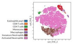
מרכז תשתיות ביורפואי מהווה מרכז ידע ומכשור טכנולוגי מתקדם החיוני לחקר מגוון שאלות ביולוגיות ורפואיות. מכשיר הCyTOF היא טכנולוגיה חדשנית, יחידה מסוגה, המאפשרת מדידה של פרמטרים רבים ברמת התא הבודד ומאפשרת כך לאפיין את תאי מערכת החיסון; פעילותם ותיפקודם.
הפרויקט המוצע: מחלות דלקתיות של דרכי האויר מתאפיינות בריבוי תאי דלקת בליחה. בברונכיאקטזיות וסיסטיק פיברוזיס קיים ריבוי נויטרופילים, אולם קיימת תת קבוצה של חולי ברונכיאקטזיות עם ריבוי אאוזינופילים בכיח. קיימות קבוצות שונות, כמו חולים במחלות דיכוי חיסוני, חולים עם מחלות דלקתיות (מפרקים או מחלות מעי דלקתיות) אשר תאי הדלקת בכיח שלהם לא אופיינו. שיטת ה CyTOF מאפשרת זיהוי תאי דלקת בכיח וחלוקתם לתת קבוצות, ואף זיהוי ציטוקינים תוך תאיים.(תמונה)
בפרוייקט המחקר המוצע נדגום תאי דלקת בכיח של מטופלים עם מחלות דרכי אויר דלקתיות כרוניות- סיסטיק פיברוזיס וברונכיאקטזיות מאטיולוגיות שונות, ונבצע אנליזה בשיטת CyTOF של תאי הדלקת בכיח שלהם.
הסטודנט ישתלב באיתור המטופלים, עיבוד הדגימות וניתוח התוצאות.
כתובת מייל: michalsh@technion.ac.il; amirgrau@technion.ac.il
 אבולוציה של רשתות בקרה גנטיות
ד״ר אלה פרגר-בן נון, מחלקה : גנטיקה וביולוגיה התפתחותית
תחומי העניין של המעבדה: המעבדה עוסקת בשאלות היושבות על הצומת בין ביולוגיה התפתחותית, בקרה גנטית ואבולוציה. אנחנו חוקרים את המנגנונים הגנטיים, המולקולריים וההתפתחותיים שיוצרים את המגוון הביולוגי המדהים הקיים בטבע. באופן ספציפי יותר, אנחנו מנסים להבין כיצד שינויים גנטיים ברצפי בקרה בגנום מובילים לשינויים אבולוציוניים בין מינים קרובים של זבובים.
הפרויקט המוצע: כיצד שינויים בגנום מובילים לאבולוציה של מבנים מורכבים? אנו משתמשים בשיטות גנומיות מתקדמות של single-cell RNA sequencing, סריקות גנטיות ועריכה גנטית ע״י CRISPR/Cas9 ע״מ לזהות גנים ורצפי בקרה המשתתפים בהתפתחות ובאבולוציה של מבנים מורכבים בזבובי פירות.
כתובת מייל: pregere@technion.ac.il
 Unraveling the networks of immune stromal interactions in the tumor microenvironment
PI – Ari Glasner, Department of Immunology
Lab site – https://glasner.net.technion.ac.il
Few cancer patients benefit from immunotherapy. Even then, tumor evasion mechanisms disrupt treatments, not only in tumor cells, but also in the stromal compartment of the tumor microenvironment (TME), often dismissed as a prospective intervention target. Insight into immune-tumor interactions is therefore critical to develop new treatments. Our lab investigates immune and stromal cell interactions in the TME, mapping the roles and pathways of each active cellular element, including multilevel interactions that occur before and after therapeutic intervention. We experiment on mouse and human tissues to test and validate potential candidates for targeted rational combination therapy, Our findings will potentially provide a framework to better understand tumor immunity dynamics, thus leading to novel therapeutic strategies.
The proposed project – Study novel candidates for combination therapy
The project offers an opportunity to work with a diverse range of cutting-edge techniques to investigate novel candidates for combination therapy. Students will gain hands-on experience in molecular and biochemical methods, functional assays, and advanced flow cytometry. The work will involve cell cultures, mouse and human tissue slice models, and in vivo assays.
Email – ari.glasner@technion.ac.il
The Molecular Machines laboratory
Associate Professor Oded Lewinson.
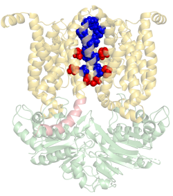
Our lab is interested in how molecular machines are built and how they work.
We are especially interested in molecular machines that are important for cancer
multidrug resistance and for bacterial pathogenesis. We harness
this knowledge to identify new targets for drug development.
For example, in 2018 our lab received the NATO Prize for Peace and Security
following our discovery that zinc can be used as a highly effective inhibitor of an essential
virulence factor of Anthrax (shown here on the right).
Email: lewinson@technion.ac.il
The Benisty Lab for Computational Neuroscience
PI- Hadas Benisty – Benisty Lab
Dept. Neuroscience
We formulate fundamental biological questions about the way the brain encodes behavior
into mathematical frameworks of interpretable models. We analyze various types of imaging
data to explore dynamics of neuronal activity and network configuration at different scales.
Computationally orientated students are welcome to apply.
 The lab of computational cancer genomics
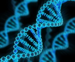
As opposed to traditional “wet” labs, much of the biological and medical research today is done computationally in what we call “dry” labs. Such research is enabled by large amount of clinical and molecular data collected around the world by the scientific community. Our lab addresses emerging challenges in computational cancer genomics by developing and applying innovative computational methods for cancer data analysis. We work closely with clinicians and experimentalists in Israel and around the world to turn data into knowledge and advance cancer treatment and prevention. One of our main interests is in predicting cancer patient response to immunotherapy using genomic, transcriptomic and metabolic data.
To join our efforts the ideal candidate should have programming skills and some level of computational background.
Email: kyizhak@technion.ac.il
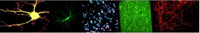
Mechanisms for life long memories
Prof. Jackie Schiller, the Cortical Computation Lab
jackie@technion.ac.il
In my lab, we study the mechanisms for lifelong learning and storage of memories.
Throughout our life, we constantly learn and memorize motor, cognitive and emotional
experiences while retaining past memories. How neurons in the brain do that?
What goes wrong in pathophysiological diseases such as autism? We use mice as
a model system to study how neurons in the motor cortex learn multiple tasks without
interfering with previously learnt tasks. We identify cell types using morphology and genetic
methods, and we study these cells and their parts, dendrites, with unprecedented resolution.
Toward this end, we use behavioral training methods combined with state of the art two-photon imaging,
viral, genetic and artificial intelligence methods.
Muscle and Nuclear Mechanobiology
The newly established Amiad-Pavlov lab is seeking curious students
for an interdisciplinary research project spanning cardiac muscle physiology,
biophysics and cell biology. We use advanced imaging approach to study
how mechanical signals dynamically regulate nuclear structure
and function in the highly contractile muscle.
For more details contact: https://md.technion.ac.il/daria-amiad-pavlov-lab/
Email: daria.amiad@technion.ac.il
 The Ubiquitin-Proteasome-System and Heart Diseases
Professor Ofer Binah. Email: binah@technion.ac.il
Department of Physiology, Biophysics and System Biology
The Research at the Laboratory
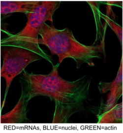
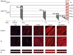
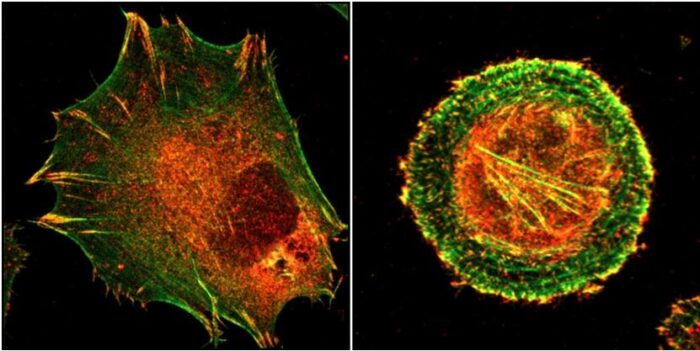
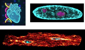
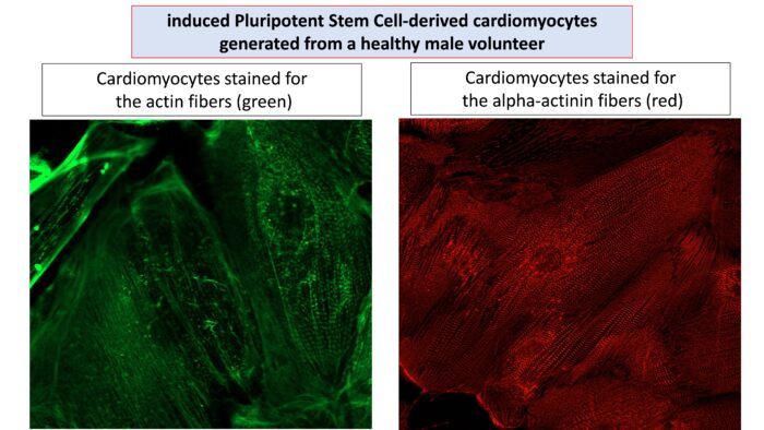
We are using induced Pluripotent Stem Cell-derived cardiomyocytes (iPSC-CMs) generated from patients suffering from inherited cardiac diseases, such as cardiomyopathies, mainly focusing on Duchenne Muscular Dystrophy (DMD) – a lethal disease with no cure, causing death at the late 20’s early 30’s. The picture illustrated iPSC-CMs generated from a healthy male volunteer. The cardiomyocytes are stained for the contractile filaments actin (left panel, green) and alpha-actinin (right panel, red).
The Ubiquitin-Proteasome System (UPS) discovered by Professors Hershko and Ciechanover (the Rappaport Faculty of Medicine, Technion) and Professor Irwin Rose (USA), is the major proteolytic cell system, which functions as a highly selective and closely regulated degradation machine, and is responsible for the degradation of most cellular proteins, native or misfolded. The UPS is especially important to the heart (with very limited self-renewal capacity), as cardiomyocytes are particularly prone to protein damage due to constant mechanical and metabolic stress. Importantly, UPS impairments have been detected in cardiomyopathies, heart failure, arrhythmias, myocardial ischemia and hypertrophy. Accordingly, our goal is to investigate the contribution of UPS dysregulation to heart dysfunction in these diseases. Importantly, discovering the discrete UPS aberrations in heart diseases can constitute the basis for developing novel therapies for alleviating the pathologies associated by heart diseases, causing massive morbidity and mortality worldwide.
Curious and scientifically motivated students are invited to discover to amazing world of induced Pluripotent Stem Cell-derived cardiomyocytes, the Ubiquitin-Proteasome System and its contribution to devastating heart diseases
Neuro-AI lab
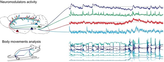
Our lab develops understanding and practical tools to study neural dynamics and learning. For example, we analyze neuromodulator dynamics (dopamine and others) to clarify how they bias actions and shape learning, build real-time interfaces to read out neural activity for novel closed-loop experiments, and develop neural network models of brain computation (RNNs, LLMs, and related approaches) to connect theory, data, and mechanism. If you’re interested in any of these, or similar research avenues, email me! stefano@technion.ac.il
Developing novel neuro-modulation treatments for epilepsy and Parkinson’ disaese
Yitzhak Schiller MD PhD
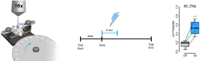 My lab uses advanced imaging and electrophysiological recordings combined with opto- and chemogenetic neuro-modulation
techniques to study the pathophysiology of two network diseases of the brain, epilepsy and Parkinson’s disease, and develop new
neuro-modulation treatment modalities to treat these diseases.
The specific proposal for the program is to join a project on opto-DBS for treating Parkinson’s disease.
In this project we attempt to improve motor performance in a Parkinsonian mouse by cell-type specific optogenetic
stimulation of different nuclei within the basal ganglia.
Cell type specific optogenetic stimulation in Parkinsonian mice
Genetics and Developmental Biology
Laboratory of Prof. Tom Schultheiss
Formation of the Ventral Body Wall in the Developing Embryo
Fields of Interest of the Laboratory: The lab investigates basic questions in embryonic development, using a broad range of molecular, microscopic, and biophysical approaches, and using the chicken embryos as a model system. We currently focus on the field of morphogenesis: how tissues, organs, and the embryo as a whole arrange their cells into specific shapes and structures.
 Description of Project: The chicken embryo (as well as the human) starts out as a flat disk of cells. This disk folds in order to form a closed gut tube (digestive system) and the ventral side of the body (the belly side). Problems in the folding process are a common cause of birth defects of the intestines and the body wall. The student will join an ongoing project that investigates the mechanisms of body wall folding, using a variety of molecular, microscopic, and biophysical approaches. The project is particularly suitable for students who are comfortable with physics and mechanics, and are interested in potentially working at the interface of biophysics and biology.
Email: tschulth@technion.ac.il
Neurotransmitters and their precursors in the regulation of neurodevelopment and behavior
Prof. Herman Wolosker
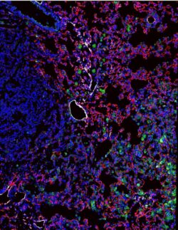
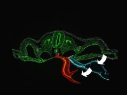
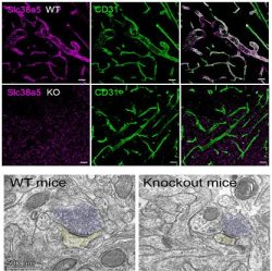 The top panel shows the presence of a new
amino acid transporter (purple) at the blood
vessels of the blood-brain barrier.
The second panel shows brains of mice in
which this transporter was removed.
The third panel shows smaller synapses
(fewer synaptic vesicles – blue and smaller postsynaptic areas – yellow)
in the knockout mice, which have smaller brains,
altered behavior and developmental problems caused
by the altered amino acid transport across the blood-brain barrier.
Lab Pi: Prof. Herman Wolosker– Department of Biochemistry, e-mail:hwoloske@technion.ac.il
Our laboratory is interested in novel neuromodulators and mechanisms that influence the formation of new neurons (neurogenesis) and synapses in the brain. Some of the genes we work are involved in Microcephaly and mental disability in children.
The project will characterize the neurochemistry, behavior, and metabolism of new mouse models that we have developed and expect to exhibit altered brain development.
Development of genetically encoded toxins for preventing neuronal degeneration
PI- Shai Berlin- Berlin Lab
Dept. Neuroscience
We focus on building tools and methods to non-invasively study and manipulate brain function at various scales: from whole brain imaging to manipulations of specific ion channels at the sub-cellular level.
Join Us!!